Aim: Genetically modified pigs have emerged as a potential source for organ transplantation. Developing effective crossmatch methods to detect humoral alloimmunity is crucial for successful xenotransplantation. In this study, we conducted flow cytometric xenocrossmatch using sera from HLA-sensitized patients, non-human primates (NHPs) with islet xenotransplants, and porcine peripheral blood mononuclear cells (PBMCs).
Method: Donor PBMCs were isolated from heparinized whole blood collected from Pigs (WT and QKO; GGTA1, CMAH, β4galNT2, iGb3s genes knock-out) using density gradient centrifugation. Human sera were obtained from patients who underwent HLA antibody testing using single-antigen Luminex bead assay (SA). 2.5x10^5 PBMC and 50 µl serum were incubated with fluorochrome-conjugated anti-porcine CD3 epsilon, CD21 and anti-human IgG, IgM antibodies. Acquisition and analysis of flow crossmatch results were performed on a Cytek spectral flowcytometer. Median fluorescent intensity (MFI) values and MFI ratios (NHP; MFI after transplantation/ MFI before transplantation, Human sera; test MFI / negative control MFI) were compared across sera to quantify antibody levels.
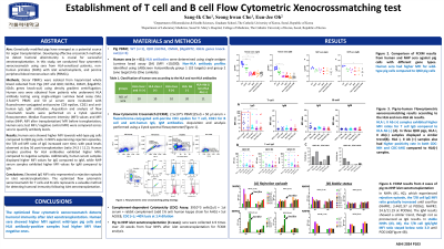
Results: Human sera showed higher MFI towards wild-type pig cells compared to QKO pig cells. In NHPs experiencing rejection episodes, the T/B cell MFI ratio of IgG increased over time, with peak levels observed at day 56 post-transplantation (ratio 24.3 / 11.2). Samples positive for HLA antibodies exhibited higher MFIs compared to negative samples. Additionally, human serum samples displayed higher MFI values for IgG compared to IgM, while NHP serum samples exhibited higher MFI values for IgM compared to IgG.
Conclusion: Elevated IgG MFI ratio represented a rejection episode in islet xenotransplantation. The optimized flow cytometric xenocrossmatch for T cells and B cells represents a valuable method for detecting humoral immunity following islet xenotransplantation.